

News Archive
Higher resolution achieved through the combination of light sheet microscopy with SIM
Mai 2017. The prime reason to use fluorescence light microscopy is the observation of live, thick, multicellular, and 3D specimens under close to natural conditions. Thus, one has to work with specimens that are mounted in an aqueous medium and not attached to or established close to a coverslip. In addition, the main issues in fluorescence microscopy, fluorophore bleaching, endogenous organic molecule phototoxicity, and solar-level intensities have to be addressed. Light-sheet-based fluorescence microscopy (LSFM) has emerged as one of the most valuable novel tools in developmental biology, plant biology, and 3D cell biology.
LSFM features optical sectioning in the excitation process. It minimizes fluorophore bleaching as well as phototoxic effects and provides a true axial resolution. The detection path resembles properties of conventional fluorescence microscopy. Structured illumination microscopy (SIM) is attractive for superresolution because of its moderate excitation intensity, high acquisition speed, and compatibility with all fluorophores. A team of scientists at Goethe University Frankfurt introduced SIM to LSFM because the combination pushes the lateral resolution to the physical limit of linear SIM. The instrument requires three objective lenses and relies on methods to control two counterpropagating coherent light sheets that generate excitation patterns in the focal plane of the detection lens. SIM patterns with the finest line spacing in the far field become available along multiple orientations. Flexible control of rotation, frequency, and phase shift of the perfectly modulated light sheet are demonstrated. Images of beads prove a near-isotropic lateral resolution of sub-100 nm. Images of yeast endoplasmic reticulum show that coherent structured illumination (csi) LSFM performs with physiologically relevant specimens. Whereas SIM is restricted to flat specimens and surfaces, csiLSFM developed by Bo-Jui Chang, Victor Perez Meza and Ernst Stelzer operates deep inside three-dimensional specimens. 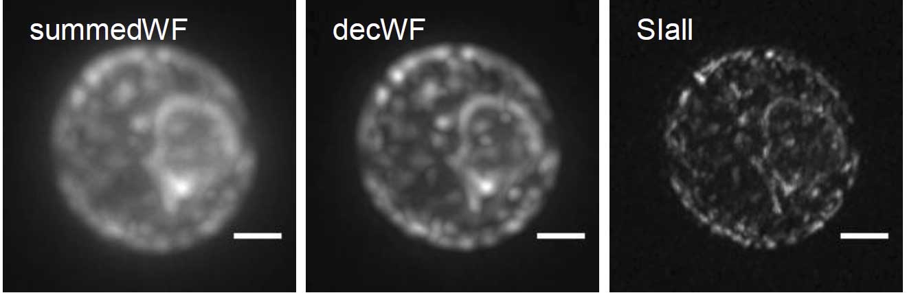
Figure: living yeast cells in agarose. From left to right: conventional fluorescence, conventional edited and csiLSFM. The bar has a length of 1 µm. (copyright Stelzer group, Goethe University Frankfurt)
Contact:
Ernst H. K. Stelzer, Institute of Cell Biology and Neuroscience, Buchmann Institute for Molecular Life Sciences, Riedberg Campus, Goethe University Frankfurt, Frankfurt am Main, Germany, Tel.: +49 (69) 798 42547/42545, ernst.stelzer@physikalischebiologie.de
Publication:
Chang B-J, Perez Meza VD, Stelzer EHK (2017) csiLSFM combines light-sheet fluorescence microscopy and coherent structured illumination for a lateral resolution below 100 nm. Proceedings of the National Academy of Sciences of the USA, published online 24 April 2017. http://dx.doi.org/10.1073/pnas.1609278114
Cluster of Excellence Macromolecular Complexes, Frankfurt am Main, Germany