News Archive
Water emerges as key player in the opening of a membrane channel important in optogenetics
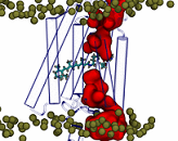
 March 2018. Channelrhodopsin-2 is a light-activated membrane ion channel that is widely used in biotechnological applications and optogenetics. Nonetheless, key elements of its structure, photocycle, and mechanism of action remain unresolved. Scientists at the Max Planck Institute of Biophysics in Frankfurt am Main applied computational modeling and over 14 µs of molecular dynamics simulation to explore the molecular mechanism of photoactivated channel opening. Water emerged as a central player that lubricates the motions of gate residues and opens a water permeable preopen pore. By resolving the early functional dynamics of ChR2, their work also provides a foundation for the design of new variants for optogenetics applications.
Channelrhodopsins (ChRs) are proteins that have revolutionized neurobiology. They were first isolated in a green alga, where they function as sensory photoreceptors for phototaxis. Their seven transmembrane helices anchor a retinal molecule. The retinal is covalently attached to the protein via a Schiff base linkage. Their function as light-activated ion channels makes ChRs unique in the rhodopsin family. Light absorption triggers a trans-to-cis isomerization of the C13−C14 double bond of the retinal chromophore. The strained 13-cis retinal moiety then induces conformational changes in the protein that open a channel through which ions can pass.
Beyond the study of neuronal circuits, ChRs are also explored as treatments of neurological disorders such as Parkinson disease, for the restoration of light sensitivity and visual capabilities, and for a range of biotechnological applications. Channelrhodopsin-2 (ChR2) in particular has found wide application thanks to its good expression in the neurons of many organisms.
Albert Ardevol and Gerhard Hummer explored the structural dynamics in the early photocycle leading to channel opening by classical (MM) and quantum mechanical (QM) molecular simulations. With QM/MM simulations, they generated a protein-adapted force field for the retinal chromophore, which they validated against absorption spectra. In a 4-µs MM simulation of a dark-adapted ChR2 dimer, water entered the vestibules of the closed channel. Retinal all-trans to 13-cis isomerization, simulated with metadynamics, triggered a major restructuring of the charge cluster forming the channel gate. On a microsecond time scale, water penetrated the gate to form a membrane-spanning preopen pore between helices H1, H2, H3, and H7. This influx of water into an ion-impermeable preopen pore is consistent with time-resolved infrared spectroscopy and electrophysiology experiments. In the retinal 13-cis state, D253 emerged as the proton acceptor of the Schiff base. Upon proton transfer from the Schiff base to D253, modeled by QM/MM simulations, they obtained an early-M/P2390–like intermediate. Rapid rotation of the unprotonated Schiff base toward the cytosolic side effectively prevented its reprotonation from the extracellular side. From MM and QM simulations, the scientists gained detailed insight into the mechanism of ChR2 photoactivation and early events in pore formation. More...
Contact:
Gerhard Hummer and Albert Ardevol, Max Planck Institute of Biophysics, Frankfurt am Main, Germany, gerhard.hummer@biophys.mpg.de, albert.ardevol@biophys.mpg.de
Publication:
Albert Ardevol & Gerhard Hummer (2018) Retinal isomerization and water-pore formation in channelrhodopsin-2. Proceedings of the National Academy of Sciences of the USA: published online 19 March 2018. http://dx.doi.org/10.1073/pnas.1700091115
Cluster of Excellence Macromolecular Complexes, Frankfurt am Main, Germany

