News Archive
EM study resolves a key question about the functioning of ATP synthase
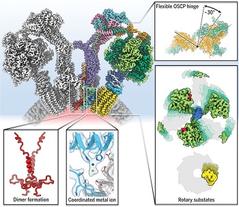
June 2019. ATP synthases located in the membrane of mitochondria generate most of the energy currency ATP, which enables higher organisms to live. Although ATP synthase complexes have been investigated for more than 50 years, several key questions remain about the way they function. One of these questions is how the stoichiometrically mismatched Fo rotor part of the ATP synthase (composed of 8 to 17 c subunits) and its three-fold symmetric F1 head are efficiently coupled. Another open question is the exact pathway taken by protons through the membrane, which has been the least well characterized part of the mechanism. A team of scientists at the Max Planck Institute of Biophysics in Frankfurt used high-resolution cryo–electron microscopy to address these questions. The scientists managed to extract 13 rotational substates of the movement of the ATP synthase which revealed that the rotation of the Fo ring and central stalk is coupled with partial rotations of the F1 head.
 Mitochondrial F1-Fo ATP synthases are macromolecular turbines that couple the location of protons across a membrane to the synthesis of ATP. Protons are translocated through the Fo subcomplex, located in the membrane, by a rotor composed of a defined number of c subunits to generate ring rotation. A central stalk is firmly anchored to the rotor ring and conveys rotary motion to the catalytic F1 subcomplex in the mitochondrial matrix, where ATP is produced by rotary catalysis. A peripheral stalk connects the two subcomplexes to prevent idle rotation of F1 with Fo.
The Frankfurt scientists used single-particle cryo–electron microscopy to characterize the structure and dynamics of a complete and active mitochondrial ATP synthase from the chlorophyll-less unicellular alga Polytomella sp. Together with data obtained by genome sequencing and mass spectrometry, the 2.7- to 2.8-Å resolution map they obtained allowed them to build a full atomic model of the 1.6 megadalton synthase complex.
The model includes the newly identified subunit ASA10, which interlinks the two ATP synthase monomers on the lumenal side of the membrane. Separation of 13 independent rotary states provided a detailed molecular description of the movements that accompany c-ring rotation.
The team found that the F1 head rotates together with the central stalk and c ring through approximately 30°, or one c subunit, at the beginning of each 120° step (movie 2). Flexible coupling of the F1 head to the Fo motor is mediated primarily by a hinge at the interdomain link of the oligomycin sensitivity–conferring protein (OSCP) subunit that joins the F1 head to the peripheral stalk. The extended two-helix bundle of the central stalk γ subunit interacts with the catch-loop region of one β subunit of the F1 head. The resulting mechanism of flexible coupling has probably been conserved in other F1-Fo ATP synthases in the course of evolution. These results, published in June 2019 in the prestigious journal Science, provide much-needed context to a wealth of published data indicating that OSCP is a hub of metabolic control in the cell.
In addition, their high-resolution map of the proton-translocating Fo complex has revealed a strong density, very likely a metal ion, ligated by two histidine residues. Recent cryo-electron microscopy studies of yeast and spinach chloroplast ATP synthase contain unannotated densities at the same position. Mutational experiments in Escherichia coli have shown that an equivalent residue is essential to proton translocation. By three-dimensional classification, the team separated two different rotational positions of the c ring and showed that the coordination environment of the metal ion changes with c-ring position. This evidence points toward a role for the metal ion in synchronizing c-ring protonation with its rotation.
Contact:
Werner Kühlbrandt, Department of Structural Biology, Max Planck Institute of Biophysics, Frankfurt/Main, Germany, werner.kuehlbrandt@biophys.mpg.de
Publication:
Bonnie J. Murphy, Niklas Klusch, Julian Langer, Deryck J. Mills, Özkan Yildiz & Werner Kühlbrandt (2019) Rotary substates of mitochondrial ATP synthase reveal the basis of flexible F1-Fo coupling. Science 364: eaaw9128. http://dx.doi.org/10.1126/science.aaw9128

