

News Archive
Functional insights into the rhodopsin activation process
February 2014. One of the most thoroughly studied G protein-coupled receptors is the mammalian visual dim-light photoreceptor rhodopsin. In collaboration with Josef Wachtveitl from Frankfurt and Judith Klein-Seetharaman from the University of Warwick, UK, a team of scientists led by Harald Schwalbe succeeded in characterizing the kinetics of its light-activation process.
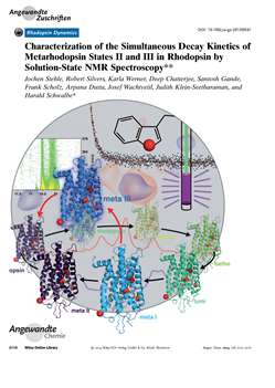 As described in their paper published in the journal Angewandte Chemie International Edition, the team found kinetic partitioning in rhodopsin decay. This included three half-lives revealing two parallel processes subsequent to rhodopsin activation that are related to the photocycle. The meta II and meta III states emerge in parallel with a relative ratio of about 3:1. Transient formation of the meta III state was confirmed by flash photolysis experiments. From analysis of the site-resolved kinetic data the scientist propose that the E2 -loop is involved in the formation of the meta III state.
In addition to providing functional insights into the rhodopsin activation process, these experiments also demonstrate that time-resolved liquid-state NMR spectroscopy can be utilized to characterize the kinetics of structural changes of membrane receptors. This represents an important advance in method development for the study of membrane protein structure and dynamics in solution. The technique thus provides a unique opportunity to link information from structural snapshots obtained by X-ray crystallography to spatially resolved kinetic information in solution.
Contact:
Harald Schwalbe
Institute of Organic Chemistry and Chemical Biology
schwalbe@em.uni-frankfurt.de
Full reference:
Stehle J, Silvers R, Werner K, Chatterjee D, Gande S, Scholz F, Dutta A, Wachtveitl J, Klein-Seetharaman J, Schwalbe H. 2014. Angew Chem Int Ed Engl., Feb 6. doi: 10.1002/anie.201309581. [Epub ahead of print]. More ...